Foot and Ankle Joint in Noida
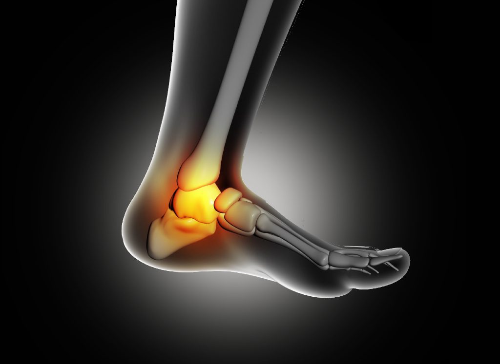
The foot has 26 individual bones connected through over 30 joints, all vital in the processes of walking and running. Even when running at a normal pace, the forces transmitted through the foot alone is approximately between three to four times a person’s body weight.
For example when someone jumps these forces can skyrocket to ten times the body weight of the individual. The ankle joint is formed by three key bones: the tibia, fibula, and talus. Moreover, the subtalar joint involves the talus, calcaneus, navicular bone, and cuboid boneset. Such joints have strong ligaments within them that support them while they move freely.
Additionally, tendons cross these joints, contributing to dynamic stabilization and preserving the integrity of joints. Any injury on this component undermines the stability and the functioning of the joint.


Special Pathologies Affecting The Ankle Joint
Achilles Tendinopathy
The Achilles tendon facilitates the transfer of leg power to the foot by connecting the calf’s triceps muscle to the heel bone. Robust tendons have the capacity to handle loads of up to 600 kg. In cases of acute injury, such as an Achilles tendon rupture, treatment may involve surgery, either through minimally invasive or open procedures. The choice between these methods depends on the nature and severity of the rupture and how quickly the injury is addressed. Following surgical intervention, functional treatment may be employed, while conservative treatment is typically reserved for specific situations where there is an increased risk associated with surgery.
For chronic Achilles tendon pain, often referred to as Achilles tendinopathy, conservative treatment is the primary approach. This treatment encompasses various physiotherapeutic interventions, along with self-administered eccentric stretching exercises. In most instances, it’s possible to continue participating in sports activities, albeit with reduced intensity and volume. Prolonged inactivity is seldom necessary and is generally not successful. An accurate diagnosis, typically based on an MRI to assess the quality and structure of the injured tendon, is crucial. Shockwave therapy is also a significant component of the treatment regimen, and PRP injections can be considered as an option.
Typical symptoms
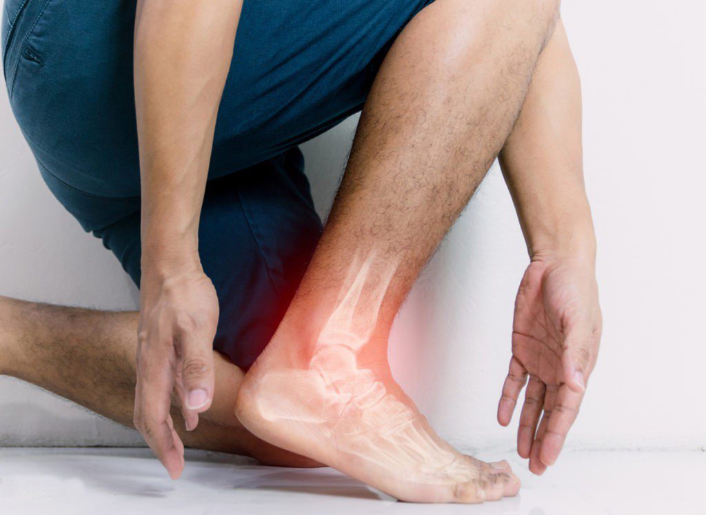
Typical symptoms of this condition include sharp pain during and after physical activity, tenderness along the tendon, or at its attachment point to the heel bone. Swelling may also be noticeable.
Surgical Intervention
In surgical procedures, whether open or arthroscopic, the patellar apex is reshaped, and any dead tissue can be removed during the same procedure.
Treatment Options
Active Strengthening: Targeted strength training is essential for fortifying overstrained muscles and preventing the recurrence of pain.
Stretching: Regularly stretching the affected muscles helps alleviate tension and supports the healing process.
Targeted Massages: Self-massage techniques can alleviate muscle stiffness and enhance blood circulation.
Anti-Inflammatory Medication: Anti-inflammatory drugs, available in forms like ointments, gels, sprays, or pills, significantly improve symptoms.
Shockwave Therapy: Concentrated pressure waves enhance local blood circulation, have a positive impact on inflammatory processes, and aid in muscle relaxation.
Insoles: Elevating the heel reduces tension in the affected tendon, leading to symptom relief.
AlterG: Utilizing an “anti-gravity treadmill” can promote healing.
Injections: For persistent pain, the injection of activated autologous blood (ACP) is often considered.”

